The Failure of Standard Orthopedic and Neurologic Tests
Part I
By Ron Eccles Jr., DC
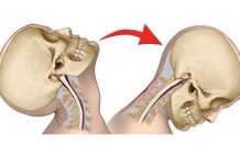
Chiropractors are regularly called upon to evaluate and treat those patients involved in motor vehicle accidents. The chiropractor often faces a significant dilemma when attempting to report findings from the standard orthopedic and neurologic tests.
On one hand the doctor realizes that the patient has been injured, however the standard orthopedic and neurologic tests that we learned in school and in postgraduate programs are not sensitive for what the patient actually suffers with. As a result of this quagmire, chiropractors often mistakenly report positive findings to substantiate necessity for care when indeed these tests should be considered negative when used appropriately.
To better explain incompatibility between the common orthopedic and neurologic tests and the whiplash patient, we must first understand the difference between radicular pain vs. referred pain.
Radicular vs. Referred Pain
Radicular pain is produced in the distribution of a nerve root as a result of some sort of mechanical compression or irritation of that root. Typically cervical radicular pain is produced from either a herniated disc or a form of entrapment secondary to degenerative change. A true radiculopathy in the cervical region will usually be found in conjunction with some sort of neurologic deficit (decreased DTRs, muscle weakness). Radicular pain syndromes are usually quite specific and localized to areas of skin innervated by a particular dermatome. “Most of the standard cervical orthopedic tests are designed to be positive only when reproduction of radiculopathy is noted.”
Referred pain is perceived in a region topographically displaced from the region of the source of the pain. The type of referred pain usually expressed in CAD trauma is “scleratogenous pain.” Scleratogenous pain is best understood by first defining the embryological relationship of tissues. Remembering back to the days in school when (or if) we studied embryology, we are reminded of somites which divide into three parts. The somite divides into the dermatome, myotome, and scleratome. From one somite, the dermatome will become a specific portion of the skin which will be innervated from that particular segment. From that same somite, muscle tissue will also form from the myotome, and again be innervated by the same segmental region. The third tissue, the scleratome, becomes connective tissue, cartilage, and bone. All tissue derived from a specific somite share one common nerve supply.
Dernatome — skin
Myotome — muscle
Scleratome — bone, cartilage, and connective tissue
Tissues Injured
The deep tissues of the cervical spine which are injured in motor vehicle accidents usually include muscles, the zygapophyseal joints,1 their joint capsules, and the surrounding connective tissue. When these tissues are lesioned, they produce a deeper, aching pain which is more difficult to localize. Their chief complaints are neck pain, head pain and pain into the upper thoracic region.
It was previously thought that scleratomes could be mapped out on charts and predictably traced according to the patient’s symptoms. It is now evident that the fields of referred pain from particular segments overlap greatly within a given individual, and the patterns exhibited by different individuals vary considerably.2 Another form of referred pain is “myotogenous pain.” The part of the somite which contributes to the formation of muscle (both apendicular and axial) is the myotome. Injury to muscles in motor vehicle accidents can produce a wide range of pain syndromes. Depending upon the level and extent of muscle injury, the pain can either be vague and difficult to localize or more pinpoint in nature. A common source of myotogenous pain comes from “trigger points.” Trigger points reproduce pain in a nondermatomal pattern and have been mapped accordingly.3
Orthopedic and Neurologic Tests
Chiropractic physicians have classically learned a group of cervical spine orthopedic tests which mostly have value assessing conditions related to radicular pain syndromes. Tests such as Bakody’s sign, Bikele’s sign, foraminal compression test, Jackson cervical compression test, maximal cervical compression test distraction test, and Spurling’s test are all designed to be sensitive for nerve root involvement.
The typical neurological examination for the cervical spine is performed in the upper extremities. The more classic tests are aimed at evaluating sensory abnormalities in the dermatomes, weakness from myotomal involvement, or abnormalities of deep tendon reflexes. While these tests prove valuable and must be performed, they are often negative and yield little information in regard to the whiplash patient.
The key to proper examination of the cervical spine lies in understanding the tissues which are involved and their nerve supply. Bogduk and his group4 have made a good case that the zygapophyseal joints of the cervical spine are a common source of pain in whiplash. Other tissues which are derived from scleratome such as ligaments and other connective tissues, along with the muscles which are derived from myotomes, seem to be the more common tissues injured in the cervical area. Most of these tissues are innervated by the dorsal ramus.
Dorsal Ramus
Peripheral nerves are made of a contribution between the dorsal and ventral roots coming off the spinal cord. These roots come together at the level of the IVF to form a mixed spinal nerve. Within a short distance of exiting the IVF, the peripheral nerve divides into four main branches. The first is a small branch, the recurrent meningeal nerve (recurrent nerve of Luschka). This nerve reenters the intervertebral foramen and ascends up and down the meninges. It provides innervation to the meninges, the intervertebral disc, and a small portion of the zygapophyseal joint capsule. Another branch of the peripheral nerve is made up of the autonomics. The autonomics will include both white and gray rami communicantes (between the levels of T1 and L2/L3), and gray rami communicantes at all levels. The largest division of the peripheral nerve is made at the ventral ramus. The ventral ramus in the cervical and upper thoracic region forms the brachial plexus. In the thoracic region, the ventral ramus becomes the intercostal nerve and in the lumbar and sacral regions, it becomes the lumbosacral plexus. The dorsal ramus is a portion of the peripheral nerve that we as chiropractors deal with more than any of the others. The dorsal ramus innervates 100 percent of the zygapophyseal joint capsule; it also supplies 100 percent of the motor contribution to the multifidus and the smaller rotator muscles. It also has contribution to the long axial muscles, the posterior ligaments and the connective tissue in this area. Tissues innervated by the dorsal ramus are among the most commonly injured by CAD trauma.
CAD Trauma
In a CAD trauma, forces usually exceed the tissue’s capability of recovery, thereby producing damage. Through damaged tissues, nerve endings become sensitized and the process of nociception begins. Nociception from spinal tissues feeds pain back to the spinal cord through the dorsal ramus.
Conclusion
As we mentioned earlier, the orthopedic and neurologic tests we typically perform are not sensitive to what produces pain in the whiplash patient. These tests, which have been designed to assess the ventral ramus of the peripheral nerve, are not usually very sensitive to the structures innervated by the dorsal ramus which are typically involved.
In the next article we will cover a better way to assess patients who are involved in motor vehicle accidents, and which tests are more sensitive for the tissues which are lesioned!
References
- Bogduk N, Marsland A. The cervical zygaphophyseal joints as a source of pain. Spine 1988; 13: 610-7.
- Bogduk N, Toomey L. Clinical Anatomy of the Lumbar Spine: Churchill Livington, 1987.
- Travell J, Simons D. Myofascial Pain and Dysfunction: The Trigger Point Manual, pg. 14.
- Bogduk N, Barnsley L et al. The prevalence of chronic zygaphophyseal joint pain after whiplash. Spine vol. 20(1) pp. 20-26, 1995


